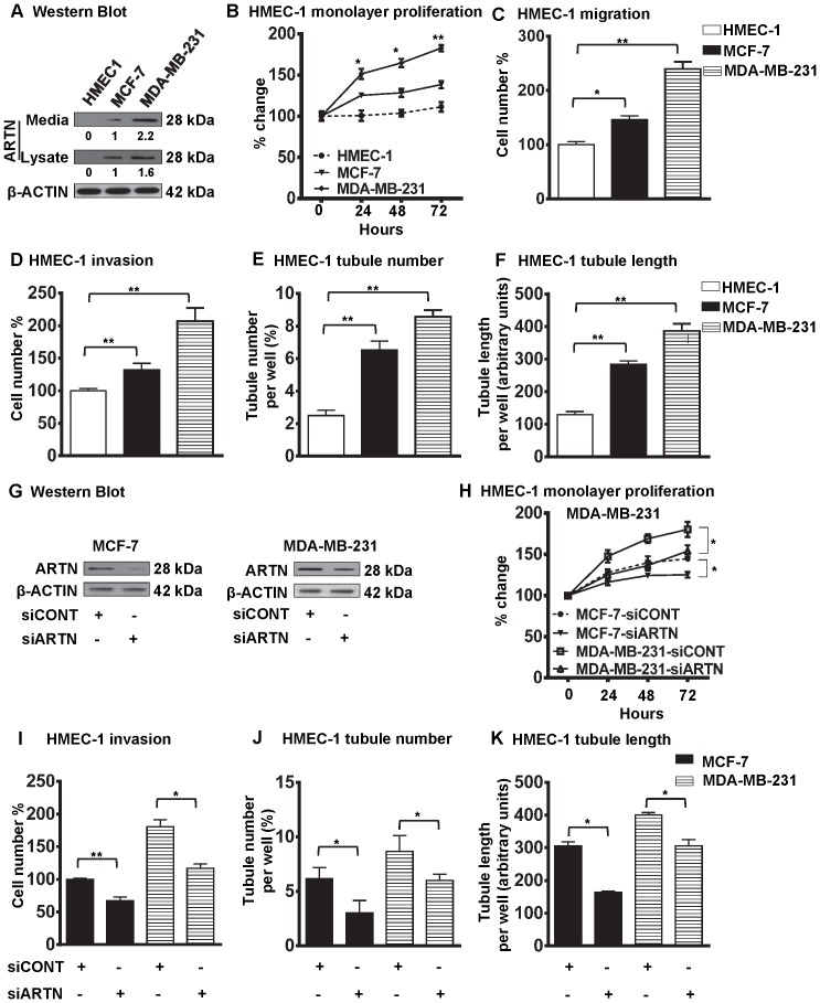
Figure 1. ARTN secreted from mammary carcinoma cells possesses a functional role in modulating endothelial cell behaviour.
(A) Western blot analysis for ARTN protein expression in HMEC-1, MCF-7 and MDA-MB-231 cells respectively. Soluble whole cell lysates or concentrated conditioned media were run on an SDS-PAGE and immunoblotted using goat anti-ARTN antibody. β-ACTIN was used as a loading control. (B) HMEC-1 monolayer proliferation. HMEC-1 total cell number after indirect co-culture with either MCF-7 or MDA-MB-231 cells in 2% serum containing media. Cell growth was measured at the indicated time points. Initial numbers of seeded cell are presented as 100%. As an internal control, HMEC-1 total cell number assay without co-culture was also performed. (C) HMEC-1 cell migration after 24 h indirect co-culture with either MCF-7 or MDA-MB-231 cells in serum free conditions. Numbers of migrated HMEC-1 cells without co-culture are presented as 100%. (D) HMEC-1 cell invasion assay after 24 h indirect co-culture with either MCF-7 or MDA-MB-231 cells in serum free conditions. Migratory or invasive cell numbers was calculated as (number of invaded cells through the inserts/total number of cells seeded) × 100. Numbers of invaded HMEC-1 cells without co-culture are presented as 100%. (E) and (F) HMEC-1 cells in vitro tube formation in matrigel after 12 h indirect co-culture with either MCF-7 or MDA-MB-231 cells in serum free conditions. HMEC-1 tubule number (E) and tubule length (F) was assessed after 12 h. Tubule number was calculated as (number of cells with tubule/total number of cells counted) × 100, whereas tubule length was calculated as an arbitrary units using ImageJ software®. As an internal control, HMEC-1 tubule number and tubule length was also assessed without co-culture. (G) Western blot analyses for ARTN in MCF-7 and MDA-MB-231 cells± siRNA to ARTN. Scrambled RNA was used as siCONT. β-ACTIN was used as loading control for cell lysates. The sizes of detected protein bands in kiloDalton (kDa) are shown on the right. (H) HMEC-1 monolayer proliferation. HMEC-1 cell numbers after indirect co-culture with either MCF-7 or MDA-MB-231 cells in 2% serum containing media ± siRNA of ARTN. Scrambled RNA was use as control. Cells growth was measured at the indicated time points. Initial numbers of seeded cell are presented as 100%. (I) HMEC-1 cell invasion assay after 24 h indirect co-culture with either MCF-7 or MDA-MB-231 cells in serum free conditions ± siRNA of ARTN. HMEC-1 tubule number (J) and tubule length (K) was assessed after indirect co-culture with either MCF-7 or MDA-MB-231 cells ± siRNA of ARTN. *, p<0.05; **, p<0.01.

