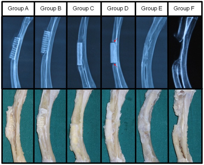
Figure 3. X-ray and gross examinations of the radius with different treatment at 12 weeks post-surgery.
X-ray examination (upper row) showed evident ingrowth of new bone tissue into the porous (Group A and B) and tubular (Group C) scaffolds. Tubular scaffolds showed better graft/bone integration than porous ones, which was comparable to the implantation of autologous bone graft (Group E). The solid scaffolds (Group D) showed the poorest osteointegration as evidenced by clear boundary line (red arrow) between the scaffold and bone tissue. Non-union healing of the bone defect in Group F (no treatment) validated that the segmental defect used in this study was the critical sized bone defect. Gross view of the radius (lower row) showed treatment with porous or tubular scaffolds and autologous bone graft exerted better bony union than solid scaffolds.

