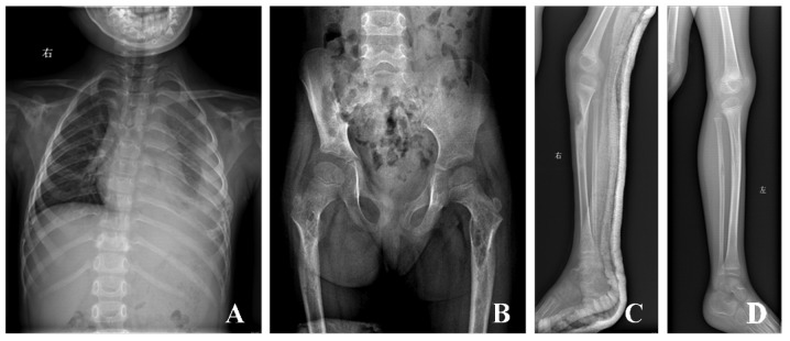
Figure 1.
(A) X-ray reveals multiple osteolysis, including bilateral clavicle, bilateral scapula, the 4th, 5th, 9th, 10th right ribs, the 2th and 6th-10th left ribs, the third, fourth and fifth lumbar vertebra, (B) right ilium, sacrum, bilateral femur, (C) right tibia, and (D) left tibia was normal as control.

