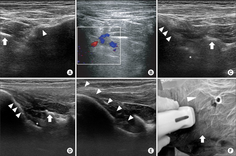
Fig. 1.
Ultrasound guided atlanto-occipital joint injection. (A) In ultrasound view of living body at the longitudinal scan below the mastoid process, C1 transverse process (arrow) and C2-3 facet joint (arrowhead) are shown. (B) Doppler examination in living body. Vertebral artery is visualized between C1 transverse process and occiput. (C) In ultrasound view of living body with a longitudinal scan at the mid-point between mastoid process and occipital protuberance, atlanto-occipital joint (asterix) is seen between C1 transverse process (arrow) and occiput (arrowhead). (D) In ultrasound view of cadaver, same as C, atlanto-occipital joint (asterix), C1 transverse process (arrow), and occiput (arrowhead) are shown. The atlanto-occipital joint is more clearly visualized than in living body. (E) In real time ultrasound guided injection, needle (arrowhead) is placed at the atlanto-occipital joint. (F) Needle insertion point is the mid-point between occipital protuberance (arrowhead) and mastoid process (arrow).

