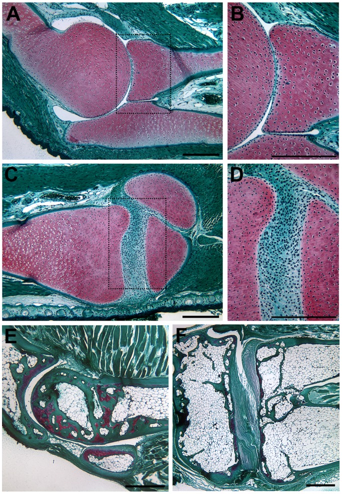
Figure 2. Joints in the forelimb and hind limb of the post-metamorphic axolotl and frog (Xenopus tropicalis).
The elbow joint (A, B) and knee joint (C, D) of a post-metamorphic axolotl (treated with thyroid hormone to induce metamorphosis) were morphological the same as in neotenous adult axolotls (compare to Fig. 1 A–D). In adult X. tropicalis, the synovial cavity of the elbow joint was acellular (E); whereas, and in the knee the synovial cavity contained fibro-cellular tissue (F). This is the same pattern as observed in axolotl elbow and knee joints (compare to Fig. 1 A–DFig. 1 A–D; Fig. 2 A–D). Tissue sections were stained with Fast Green/Safranin O/Weigert’s Iron Hematoxylin. Boxed areas in (A) and (C) are illustrated at higher magnification in (B) and (C) respectively. The conserved anatomy was confirmed by analysis of the joints in two different post-metamorphic axolotls and two individual X. tropicalis. Scale bars = 500 µm.

