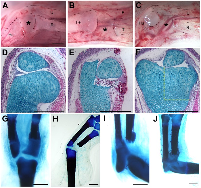
Figure 5. Regeneration of sub-critical size defects in axolotl limb joints.
A defect in the distal region of the epiphysis of the humerus (A, D–E) was regenerated (F) within 8 weeks after surgery (macroscopic view of the surgery is illustrated in A). Similarly, a 1 mm defect in the proximal epiphysis of the tibia (surgery illustrated in B) regenerated (G), as did the same size defect in the proximal radius (J; surgery illustrated in C). The regenerative response to excision of 1 mm of the proximal radius was variable. In smaller animals (8–10 cm snout to tail tip), a 1 mm defect failed to regenerate (I); however, in larger animals (12–14 cm), a 1 mm defect was regenerated within 6 weeks post-amputation (J). Defects greater than 1 mm failed to regenerate in either the radius (not illustrated) or the tibia (H). Tissue sections were stained with Alcian blue/Hematoxylin/Eosin Y (D–F). Whole-mount limbs were stained with Alcian Blue/Alizarin Red (G–J). The black dotted lines in A–C demarcate the skeletal elements that remained after the surgical defect was created (Hu, humerus; U, ulna; R, radius; Fe, femur; F, fibula; T, tibia). The yellow dotted lines in (F) indicate the region of the defect that was regenerated (compare to E). Results were confirmed by experimental replication in three animals for distal humerus excisions, seven animals for proximal tibia excisions, and eight animals for proximal radius excisions. The magnification is the same in A–C. Scale bars = 1 mm.

