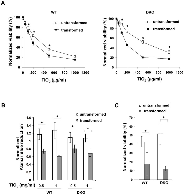
Figure 2. TiO2 nanoparticles preferentially induce cell death in transformed cells.
(A) The indicated cell lines were treated with various concentrations of P25 TiO2 nanoparticles for 24 hours, and viability of cells was measured using TOTO-3 DNA dye exclusion method. Data represent mean±S.D. of three independent experiments. * p<0.05, Student’s unpaired t test. (B) Effects of TiO2 nanoparticles on cellular metabolic activities were determined by measuring Alamar Blue fluorescence. The indicated MEF cells were treated with 0.5 mg/ml or 1 mg/ml TiO2 nanoparticles for 24 hours. The cellular reducing activities of treated cells were normalized to that of corresponding untreated cell lines. Mean±S.D. of three independent experiments are shown. * p<0.05, Student’s unpaired t test. (C) Effects of TiO2 nanoparticles on long-term cell viability were determined by clonogenicity assay. The normalized cell survival was calculated by dividing the number of wells with viable treated cells with that of untreated cells. Data represent mean±S.D. of three independent experiments. * p<0.05, Student’s unpaired t test.

