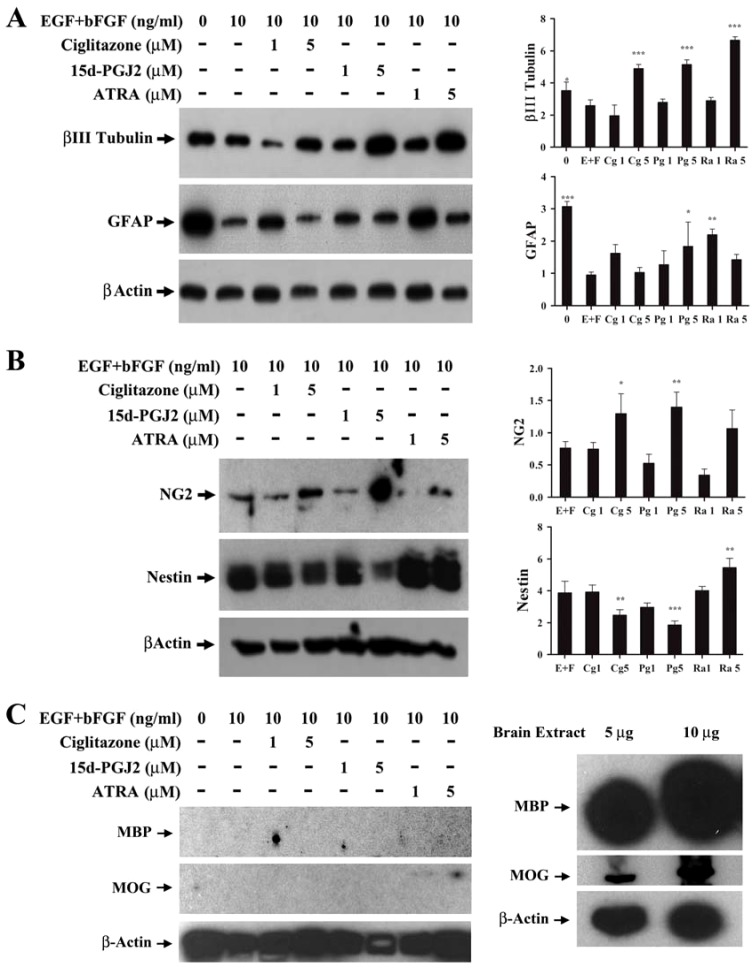
Figure 3. Modulation of stem cell and differentiation markers by PPARγ agonists in NSCs.
NSCs were cultured in NBM+B27 with EGF+bFGF in the presence of 0, 1 and 5 µM ciglitazone, 15d-PGJ2 or ATRA at 37°C for 72 h. The expression of βIII tubulin, GFAP (A), NG2, Nestin (B), MBP, MOG (C) and β-Actin was analyzed by Western blot and ECL detection system. Mouse brain extract was used as positive control (C). The relative quantities of protein bands normalized to β-Actin in the blots were determined by densitometry and presented as histograms. The values are mean±SD and the p values are expressed as *(p<0.05), **(p<0.01), and ***(p<0.001). The figure is a representative of five independent experiments.

