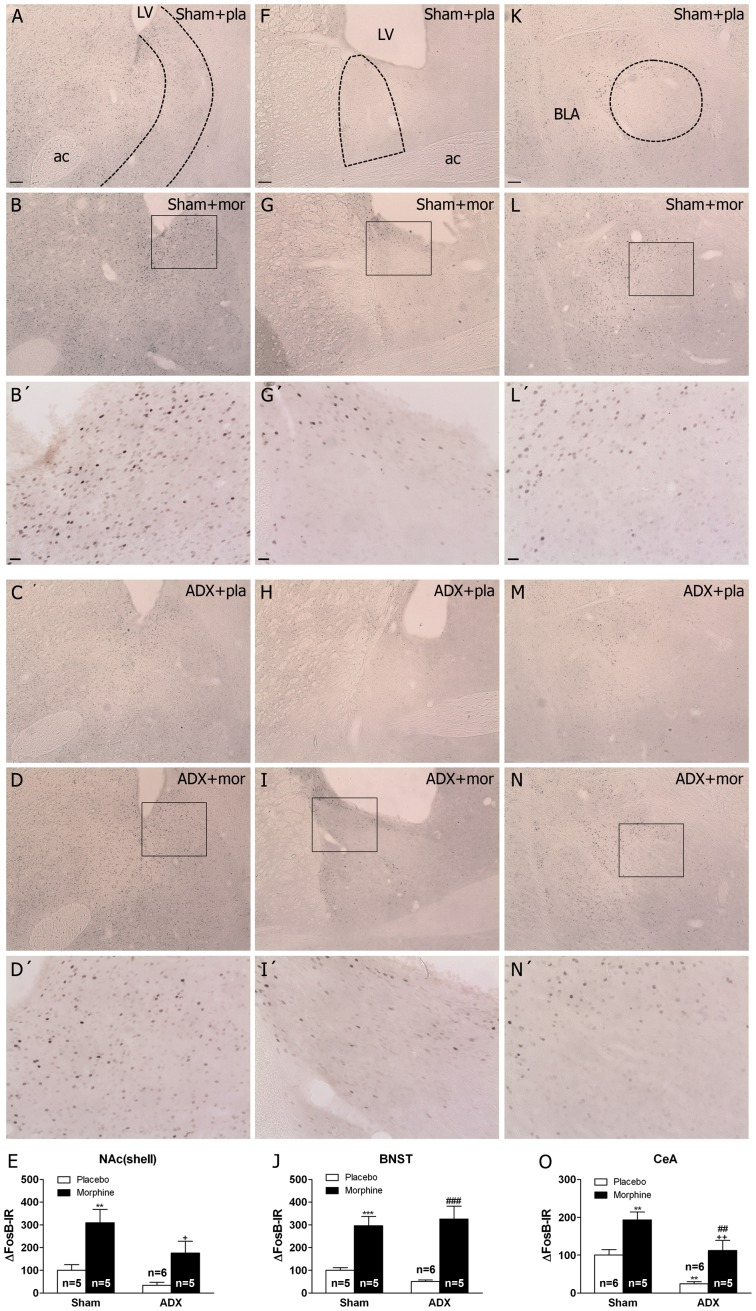
Figure 2. Adrenalectomy differentially regulates FosB/ΔFosB protein expression in the brain stress systems.
Photographs represent immunohistochemical detection of FosB/ΔFosB in the NAc(shell) (A-D), BNST (F-I) and CeA (K-N) from sham-operated and adrenalectomized (ADX) rats pretreated with placebo (pla) or morphine (mor) pellets for 10 days. B’, D’. G’, I’, L’ and N’ are high magnifications. Scale bar: 100 µm (30X, low magnification); 20 µm (100X, high magnification). LV, lateral ventricle; ac, anterior comisure; BLA, basolateral amygdala. E, J, O, quantitative analysis of FosB/ΔFosB immunoreactivity in the three nuclei from sham and ADX animal. Data correspond to the mean ± SEM (percent of control). Post hoc comparisons revealed a significant increase of FosB/ΔFosB protein expression in sham animals after chronic morphine exposure in the NAc(shell), BNST and CeA (**p<0.01; ***p<0.001 versus sham-placebo animals). In ADX-morphine dependent rats it was observed an attenuation of FosB/ΔFosB expression in the NAc(shell) and CeA compared with sham-dependent rats (+p<0.05; ++p<0.01 versus sham+morphine). In addition, ADX decreased basal FosB/ΔFosB expression in the CeA (**p<0.01 versus sham-placebo group. ##p<0.01; ###p<0.001 versus ADX+placebo).

