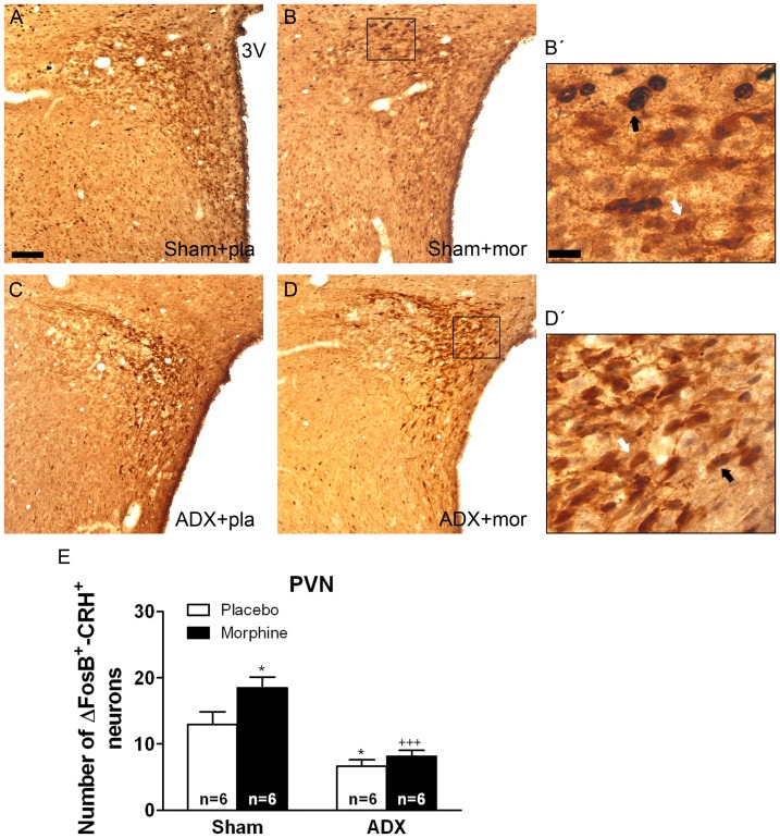
Figure 5. Adrenalectomy attenuated FosB/ΔFosB protein expression in the PVN CRH-positive neurons.
ADX and non-operated (sham) rats were made dependent on morphine (mor) for 10 days. Controls received placebo pellets (pla). Animals were perfused and the PVN was processed for double-labelled (FosB/ΔFosB and CRH) immunohistochemistry. Panels A-D show immunohistochemical detection of FosB/ΔFosB into CRH neurons after different treatments. Low and high magnifications (B’, D’) images show FosB/ΔFosB-positive (blue-black)/CRH-positive (brown) neurons (black arrow) and FosB/ΔFosB-negative/CRH-positive (white arrow) immunoreactivity. Scale bar: 100 µm (50X, low magnification); 20 µm (250X, high magnification). 3 V, third ventricle. E: quantitative analysis of FosB/ΔFosB-positive/CRH-positive neurons in the PVN. Data correspond to mean ± SEM. Post hoc test revealed a significant higher number of FosB/ΔFosB-positive nuclei into CRH immunoreactive neurons in morphine-dependent rats (*p<0.05 versus sham-placebo). ADX attenuated the increase of FosB/ΔFosB expression in CRH-positive neurons both in placebo- (*p<0.01 versus sham+placebo) and in morphine-treated animals (+++p<0.001 versus sham+morphine).

