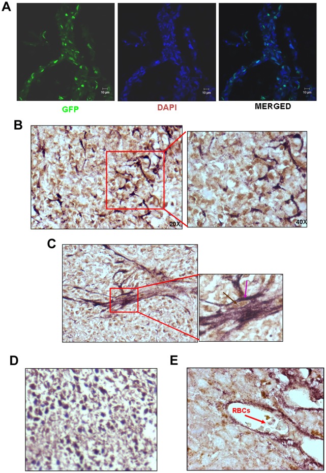
Figure 3. HT1080 tumour cells employ vasculogenic mimicry to accommodate in situ hypoxia. A.
Visualization of GFP+ tumour cells in the cryosections of the tumours formed by HT1080/WT/GFP cells showed that the tumour cells themselves form vessels that run criss-cross in the tumour mass. DAPI was used to demarcate the nuclei (bar - 10 µm) B. Double IHC experiments performed on the paraffin sections of the tumours formed by HT1080-GFP cells with anti-GFP and anti-PECAM antibodies show that the entire tumour mass was filled with GFP+ tumour cells with the vessels formed by the double positive cells. The intensity of PECAM staining was maximal in the cells forming tubes. (Original magnification 200X). A part of the image has been magnified to show the details (original magnification 400 X). C. A vessel formed by the double positive cells is shown (original magnification 200X). The inset clearly shows that the cells forming the vessels have brown nuclei (GFP – indicated by a brown arrow) and violet border (PECAM – indicated by a violet arrow) (original magnification 630 X). D. Image shows absence of PECAM staining in the tumour section of HT1080 where anti-PECAM antibody has not been applied; whereas the GFP signal is clearly seen (Original magnification is 200X). E. A cross section of the blood vessel formed by GFP-PECAM double positive cells is depicted. Presence of red blood cells in the lumen is marked by a red arrow (original magnification 630X).

