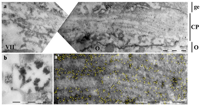
Figure 6. Fully formed contact plate immunogold stained.
Staining conditions are the same as on Fig. 5. a –contact plate of the oocyte VII final stage at low magnification. Indicated with lines at the right are: O – oocyte, CP – contact plate, ge – germinal epithelium. Scale bar –5 µm. a′ – high magnification of the part of the image on a; gold particles pseudocolored yellow. Scale bar–2 µm. b – type 2 granule presumably in the process of excretion (not stained). Scale bar – 2 µm.

