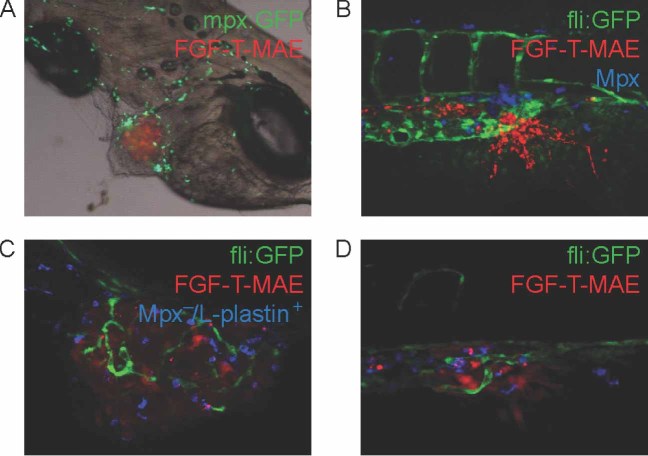
Figure 4.
Myeloid cells associated with tumour cells at the localized tumour and micrometastasis sites. (A) Neutrophils [green; Tg(mpx:GFP) ] associated with FGF-T-MAE tumour cells (red) at the localized tumour growth site. (B) Neutrophils (blue; Mpx+ histochemical staining) associated with the tumour cells (red) at the micrometastasis site. (C, D) Macrophages (blue; Mpx−/L-plastin+) associated with the tumour cells (red) at the localized tumour (C) and micrometastasis (D) sites. Data are representative images of > 10 independent, reproducible experiments (each n > 30). Image in (A) was acquired using a Leica MZ16FA stereo microscope (bright-field/fluorescence overlay image); those in (B–D) were acquired using a Leica TCS SPE confocal microscope with a × 20 dry objective

