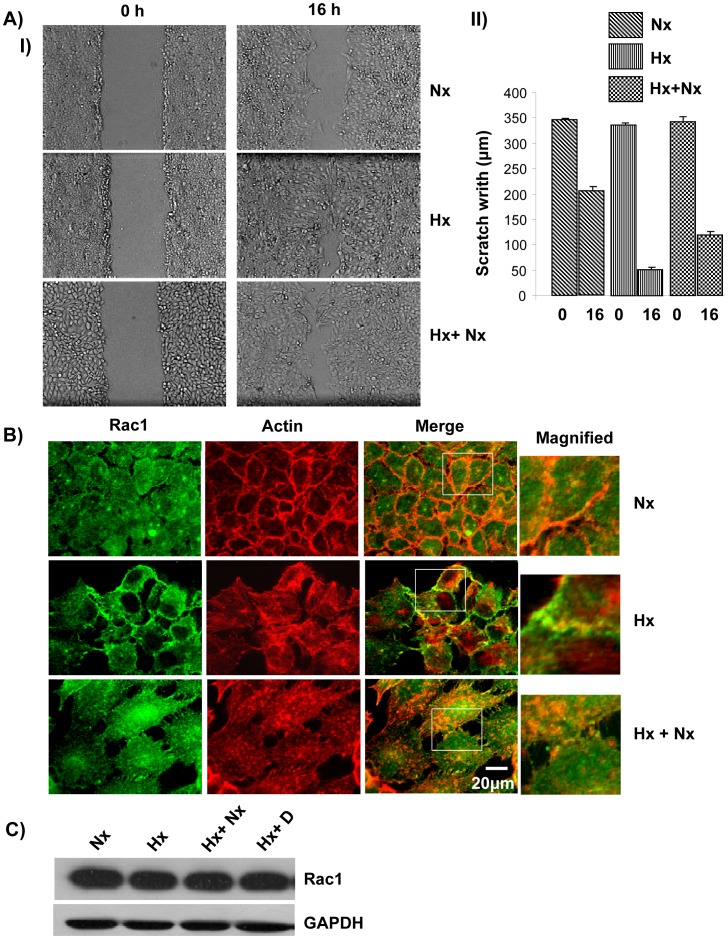
Figure 2. Growth under hypoxic conditions led to increased cell motility and re-localization of Rac1.
A) Cells grown under hypoxic conditions displayed increased cell motility. (I) A431 cells grown under normoxia, hypoxia or hypoxia+normoxia were seeded in a 6 well dish and allowed to form a monolayer. A wound was generated and imaged at 0 h and at 16 h. (II) The width of the scratch was quantified at 10 points along the scratch and averaged. The experiment was repeated three times. B) Rac1 re-localized to the plasma membrane under hypoxic condition. A431 cells grown in normoxia, hypoxia or hypoxia+normoxia conditions were probed with anti-Rac1 followed by labeled secondary antibody (Green). The actin cytoskeleton was visualized using Alexa568-Phalloidin (Red). C) Hypoxia does not lead to increased expression of Rac1. Cell lysate from A431 cells grown under normoxia (Nx), hypoxia (Hx), hypoxia followed by normoxia (Hx+Nx) or hypoxia+Cetuximab (Hx+D) were analyzed by immunoblotting with anti-Rac1 or anti-GAPDH primary antibody.

