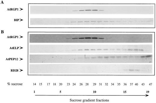
Figure 5.
Membrane localization of AtRGP1. Equilibrium-density gradients were used to fractionate A. thaliana total membrane preparations from suspension-cultured cell protoplasts. One-twentieth (100 μL) of equal-volume fractions was separated by SDS-PAGE and transferred to nitrocellulose membranes. A, Fractionation of AtRGP1 was determined by immunoblot analysis and compared with that of the membrane marker BiP (ER). Because of the presence of AtRGP1 and BiP in the same fractions, similar gradients were analyzed in the presence of Mg+2 and showed that BiP shifted to a denser part of the gradient, but AtRGP1 did not (data not shown). B, AtRGP1 fractionation was also compared with that of other known membrane markers, AtELP, AtPEP12p, and R28.

