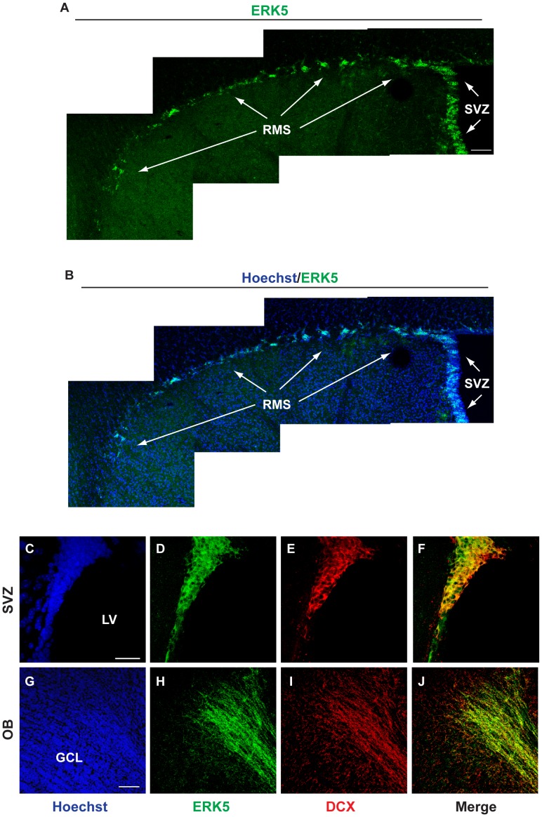
Figure 1. ERK5 is expressed along the entire SVZ/RMS/OB axis.
A, B) ERK5 protein (green) was identified by IHC along the SVZ and RMS of adult mouse brain. Hoechst staining (blue) was used to identify all cell nuclei. Scale bar in A represents 100 µm and applies to B. C–J) ERK5 co-localizes with DCX+ cells (red) along the lateral ventricles (LV) and in the center of the granular cell layer (GCL) of the OB where adult born neurons exit the RMS. Scale bar in C represents 25 µm and applies to D–F, while scale bar in G represents 100 µm and applies to H–J. Three individual mouse brains were analyzed for ERK5 expression along the SVZ/RMS/OB axis.

