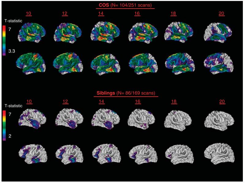
Figure 2.
Cortical gray matter (GM) deficits across age 10–20 years in patients with childhood-onset schizophrenia (COS; upper panel) and their healthy siblings compared with matched healthy controls. Statistically significant gray matter cortical thickness deficits, using mixed-effect models across over 40 000 cortical points in each hemisphere, are represented by colors corresponding to t-values shown in the scale bars. Figure 2 adapted from the research of Greenstein et al.47 (upper panel), Gogtay et al.52 (lower panel) and Mattai et al.53 (lower panel).

