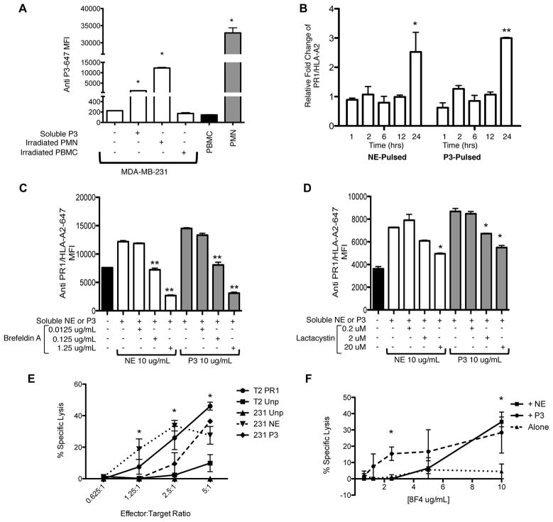
Figure 4.
Uptake of P3 and cross-presentation of P3 and NE increases breast cancer susceptibility to killing by PR1-CTLs and anti-PR1/HLA-A2. A, MDA-MB-231 breast cancer cells were incubated with soluble P3, irradiated PMNs or PBMC for 4 hours. Cells were permeabilized, stained with anti-P3 antibody and analyzed by flow cytometry. For cell-associated uptake, light scatter seen on flow cytometry provided a clear distinction between PBMC, PMN and MDA-MB-231 cells. PBMC and PMN cells alone were used as negative and positive controls, respectively. ANOVA followed by Tukey test was performed using Prism 5.0 software (*P<0.05). Data are means ± SEM from duplicate experiments. B, MDA-MB-231 breast cancer cells were cultured with soluble P3 or NE (10 μg/mL) at increasing time-points and then analyzed for expression of PR1/HLA-A2. Mean ± SEM fold increase of the median fluorescence intensity (MFI) of PR1/HLA-A2 vs. unpulsed cells is shown from duplicate experiments. ANOVA followed by Tukey test was performed using Prism 5.0 software (*P=0.01); (**P<0.0001). C and D, MDA-MB-231 cells were cultured for 24 hours in media containing NE or P3 (10 μg/mL) and the antigen presentation inhibitors brefeldin A or lactacystin. Cells were then analyzed for expression of PR1/HLA-A2. Mean ± SEM of the median fluorescence intensity (MFI) of PR1/HLA-A2 is shown from duplicate wells of a representative experiment. ANOVA followed by Tukey test were performed using Prism 5.0 software (*P<0.01); (**P<0.0001). E, MDA-MB-231 cells were cultured overnight in media containing P3 or NE (10 μg/mL), loaded with calcein-AM, and then co-cultured with PR1-CTLs for 4 hours. Cytotoxicity was determined by measuring released calcein-AM. NE- or P3-pulsed cells show higher killing vs. unpulsed MDA-MB-231 cells. PR1-pulsed and unpulsed T2 cells were used as positive and negative controls, respectively. Data are means ± SEM from duplicate wells from a representative experiment. F, MDA-MB-231 cells were cultured with NE (10 μg/mL) or P3 (10 μg/mL) for 24 hours. Cells were then incubated with anti-PR1/HLA-A2 (8F4) antibody for 60 minutes and then complement was added. Complement-dependent cytotoxicity was measured using calcein-AM release and shows specific killing of NE- or P3-pulsed MDA-MB-231 cells by 8F4 antibody. Cytotoxicity data are means ± SEM from duplicate wells from a representative experiment. Unpaired t test was performed using Prism 5.0 software (*P<0.05). Abbreviations; PMN, polymorphonuclear cells; PBMC, peripheral blood mononuclear cells.

