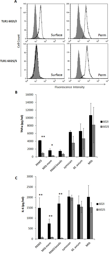
Figure 1. TLR1 602S/S macrophages are activated by whole bacteria but not soluble agonists.
A. Representative surface (left column) and permeabilized (right column, perm) staining of TLR1 in primary human monocyte-derived macrophages from a TLR1 602I (top row) and TLR1 602S (bottom row) homozygous blood donor is shown (filled histogram; isotype control, black histogram; TLR1). Primary human monocyte-derived macrophages from venous blood donors of the various TLR1 602 genotypes were stimulated with agonists, as indicated, for 24 hours and secretion of TNFα (B) and IL-6 (C) was measured from culture supernatants by ELISA (black bars; TLR1 602I/I or TLR1 602I/S donors, gray bars; TLR1 602S/S donors). Error bars represent the standard deviation of at least three donors. Asterisks denote significant differences between TLR1 602I-expressing cells versus TLR1 602S/S cells (*p<0.05, **p<0.005). (PAM3; PAM3CSK4Mtb mem; M. tuberculosis membrane fraction, PAM3 beads; PAM3CSK4-coated polystyrene beads, Mtb; gamma-irradiated M. tuberculosis)

