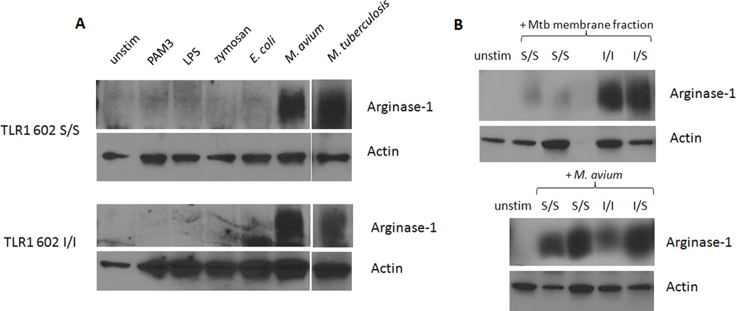
Figure 5. TLR1 602S/S monocytes resist induction of host arginase-1 when stimulated with mycobacterial membrane, but not whole mycobacteria.
A. Blood monocytes from donors of the indicated TLR1 602 genotypes were stimulated with various TLR agonists for 24 hrs followed by detection of Arginase-1 in cell lysates via Western blot. Loading controls were performed by detection of actin. B. Primary human monocytes from the indicated donors were stimulated with M. tuberculosis Mtb) membrane fraction (top), or live M. avium (bottom) for 24 hours. Levels of Arginase-1 were determined by Western blot. Relative protein loading is denoted by actin blots.

