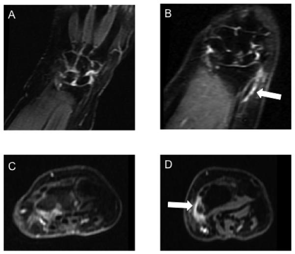
Figure 2.

MRI of the hands at baseline (A & C) and 6 months (B & D) in a patient with new onset DeQuervain’s tenosynovitis. Demonstrates abnormal increased T2 signal on coronal stir (B) and enhancement on axial post-contrast images (D) in the first extensor compartment including the right abductor pollicis longus and extensor pollicis brevis tendons
