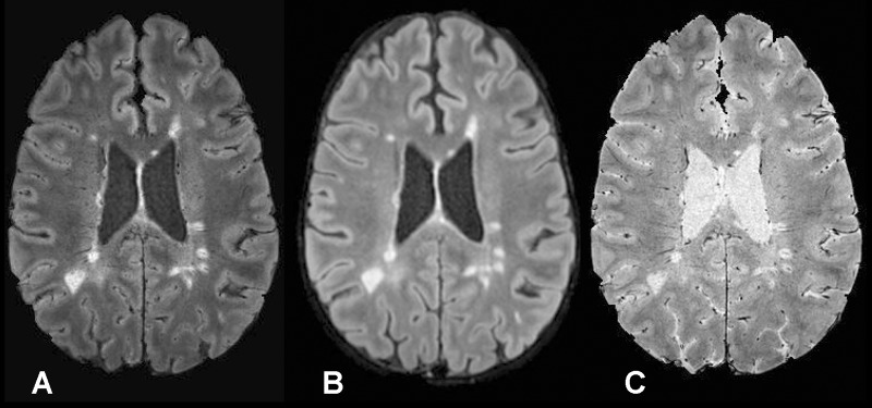
Figure 1:
A, Axial FLAIR* image (0.55 × 0.55 × 0.55 mm3 resolution), B, axial T2-weighted FLAIR image (1 × 1 × 1 mm3 resolution; repetition time, 4800 msec; echo time, 372 msec; inversion time, 1600 msec), and, C, axial T2*-weighted segEPI image (0.55 × 0.55 × 0.55 mm3 resolution; repetition time, 53 msec; echo time, 29 msec) in a 43-year-old man with relapsing-remitting MS (EDSS = 1.0, disease duration = 12 years).

