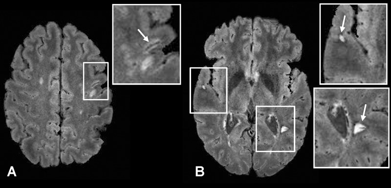
Figure 6:
Axial FLAIR* images (0.55 × 0.55 × 0.55 mm3 resolution) in a 42-year-old man with relapsing-remitting MS (EDSS = 2.5, disease duration = 3 years). A juxtacortical lesion with its central vein (arrow, A) and two lesions with hypointense rims (arrows, B) are clearly depicted. Note that these “rim” lesions also have central veins.

