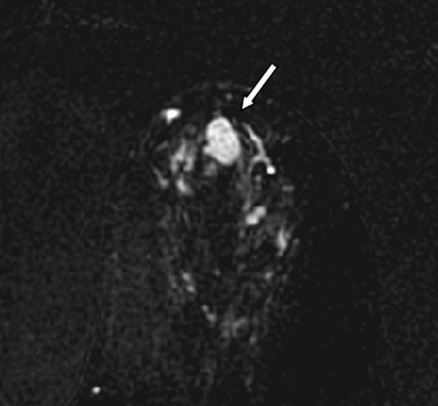
Figure 3a:

Images in 61-year-old woman with personal history of right-breast ductal carcinoma in situ. The patient underwent breast MR imaging for high-risk screening. (a) Axial dynamic contrast-enhanced initial postcontrast subtraction MR image shows 13-mm lobular heterogeneously enhancing mass (arrow) in the subareolar region of the left breast, 16 mm from the nipple. The lesion has a smooth margin and is at an anterior depth. This lesion was classified as BI-RADS category 4. On (b) an axial dynamic contrast-enhanced MR image, the lesion (arrow) shows mixed kinetics overall: 28% delayed persistent enhancement (blue), 34% delayed plateau (green), and 38% delayed washout (red). The lesion (arrow) is hypointense on (c) an axial T2-weighted MR image. The lesion (arrow) is hyperintense on (d) axial DW image and has a low ADC (mean, 1.06 × 10⁻³ mm2/sec) on (e) an ADC map. Insets in d and e = ROIs. The lesion was classified as ADH on the basis of (f) US-guided core biopsy results, which showed intraductal papilloma and ductal hyperplasia with focal atypia with no evidence of invasive carcinoma. (Hematoxylin-eosin stain; original magnification, ×200.) No evidence of carcinoma was detected at excisional biopsy.
