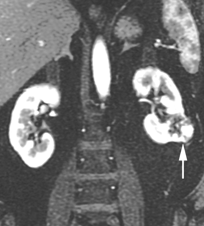
Figure 3b:

Coronal images in 68-year-old man with clear cell RCC (arrow) in lower pole of left kidney. (a) T2-weighted single-shot fast spin-echo image shows that tumor has predominantly high signal intensity relative to renal cortex. (b) T1-weighted spoiled gradient-echo image obtained during corticomedullary phase after administration of gadopentetate dimeglumine (0.1 mmol per kilogram body weight) shows that tumor exhibits avid enhancement. (c) ASL image shows heterogeneous perfusion of tumor (mean perfusion = 152.4 mL/min/100 g).
