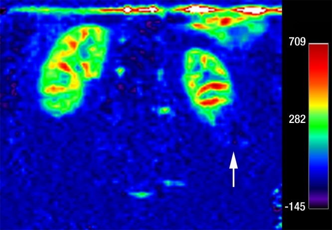
Figure 4c:

Coronal images in 64-year-old man with papillary RCC (arrow) in lower pole of left kidney. (a) T2-weighted image shows diffuse low signal intensity in mass. (b) Contrast-enhanced image obtained in the excretory phase shows that tumor has low levels of enhancement compared with renal parenchyma. This was also seen during the corticomedullary phase (not shown). (c) ASL image shows that tumor has low perfusion levels (mean perfusion = 50.0 mL/min/100 g).
