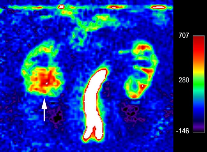
Figure 5c:

Coronal images in 61-year-old man with oncocytoma (arrow in b and c) in lower pole of right kidney. (a) T2-weighted image shows intermediate-signal-intensity tumor. (b) Image obtained in corticomedullary phase after administration of contrast material shows homogeneous tumor enhancement. (c) ASL image shows marked perfusion of entire mass (mean perfusion = 309.6 mL/min/100 g).
