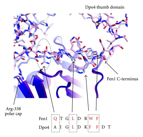
Figure 4.

Detailed comparison of the PIP-box interactions of SsoFen1 and Dpo4 with PCNA (see also Figures 2(a) and 2(b)). Fen1 and PCNA1 are shown in light and dark blue, respectively. Dpo4 and its respective PCNA1 are shown in pink and magenta. The position of the conserved glutamine residue is highlighted and the key residues in the interaction motif are indicated in red (2IZO and 3FDS, [9, 10]). Residues in the top right hand corner are part of the Dpo4 thumb domain and are proximal to the extreme C-terminus of the protein.
