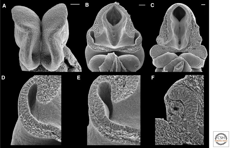
Figure 1.
Anatomy of embryonic mouse forebrain and eyes. (A) Frontal view of embryonic day (E) 8.5 forebrain, just before the eye field splits. The optic sulci are the large pits protruding from the ventral neural ectoderm. (B) Wide and (D) high magnification views of frontal sections of the E9.0 to E9.5 optic vesicle. (E) The coordinated invagination of the distal optic vesicle and the surface ectoderm, where the lens placode has thickened, begins at E9.5. (C) Wide and (F) high magnification views of frontal sections of the E10.5 optic cup and lens vesicle. The retinal pigment epithelium is the thin layer of cells proximal to the neural retina, which is dorsal to the optic stalk. The optic stalk is continuous with the ventral forebrain. The lens vesicle is distal to the neural retina. Dorsal is to the top (A–F), and proximal is to the right (D–F). Scale bars, 50 μm (A); 100 μm (B,C). (Photo from Lee Langer.)

