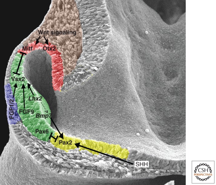
Figure 3.
Signaling networks establish boundaries in the optic vesicle. Dorsal is to the top, and distal is to the left. The optic vesicle is regionalized into prospective RPE (red, dorsal), neural retina (green, central) and optic stalk (yellow, ventral). Extracellular signals organize the optic vesicle in part through the activation of transcription factors that specify the tissue type in which they are expressed. These transcription factors cell-intrinsically regulate optic vesicle organization through mutual repression of one another. The dotted arrow indicates that early Lhx2 expression may be required for Bmp7 expression in the optic vesicle (Yun et al. 2009), but Bmp7 expression is maintained when Lhx2 is ablated specifically in the eye field (Hagglund et al. 2011). The lens placode, which expresses fibroblast growth factor (FGF) ligands important for neural retinal specification, is shown in blue.

