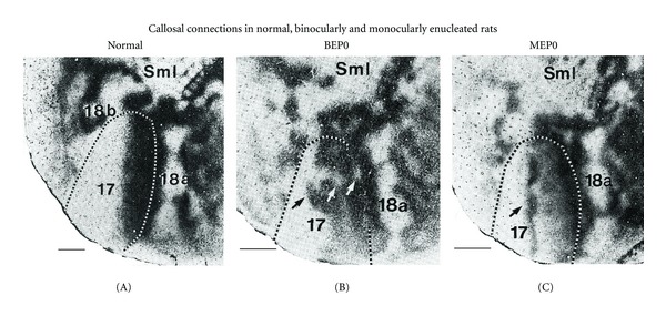
Figure 1.

Effect of binocular or monocular enucleation at birth on the pattern of rat visual callosal connections. Callosal patterns in the right hemisphere were revealed following multiple intracortical injections of HRP into the left hemisphere. Images were taken from tangential sections cut through supragranular layers of the flattened cerebral cortex. Dark areas show the distribution of labeled callosal cells and axon terminations. Segmented lines indicate the border of area 17 determined from adjacent sections stained to reveal myeloarchitectonic patterns. Lateral is right, posterior is down. (A) Callosal pattern in normally reared adult rats. Callosal cells and terminations form homogeneously labeled bands at the 17/18a border and at the lateral border of area 18a, and several narrow bands of callosal connections bridge the width of area 18a at several rostrocaudal levels. (B) Binocular enucleation increases the relative width of the callosal band at the 17/18a border and causes the appearance of discrete regions of reduced labeling within the 17/18a callosal band (white arrows) and several densely labeled tongue-like regions that extend medially from this band well into area 17 (black arrow). (C) In rats monocularly enucleated at birth, the most prominent anomaly develops in the hemisphere ipsilateral to the remaining eye, where an anomalous, dense band of callosal connections runs rostrocaudally through the center of area 17 (arrow). Periodic fluctuations in the density of labeling along the length of this extra band give it a beaded appearance. SmI = Somatosensory cortex. Scale bars = 1.0 mm. Adapted from Olavarria et al. [6].
