
Figure 2.
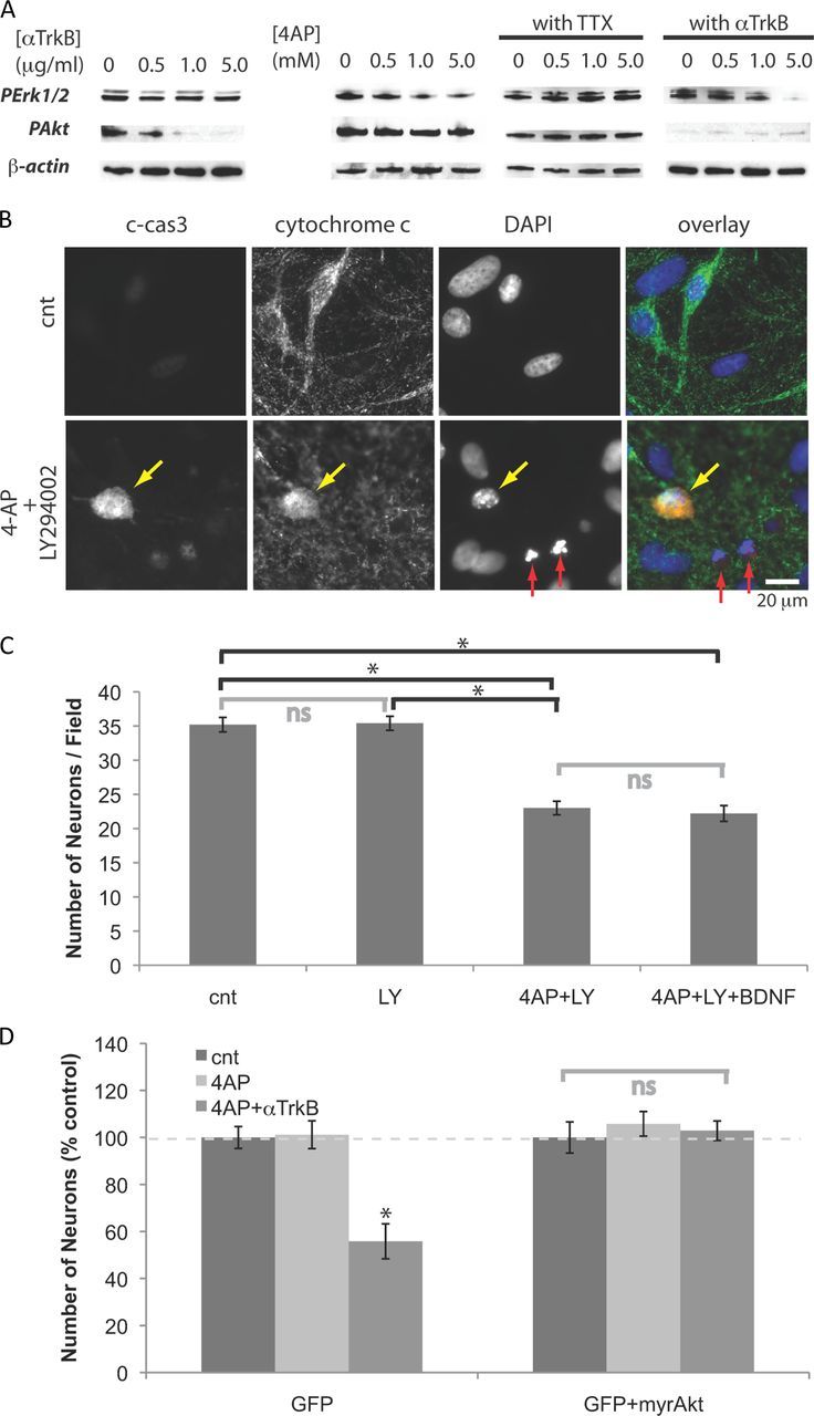
Neurons become dependent on Akt signaling when treated with 4-AP. A, Western blot analyses. Neurons were treated for 18 h with 5 mm 4-AP, and/or 5 μg/ml anti-TrkB antibody, or 1 mm TTX. B, Immunostaining for c-cas3 and cytochrome c, and DAPI staining. Neurons were treated for 2 d (from DIV12 to DIV14) with 5.0 mm 4-AP and/or 25 μm LY294002. The arrows indicate apoptotic neurons with (yellow) or without (red) activation of caspase 3. C, Numbers of neurons after 2 d treatments. Five millimolar 4-AP, 25 μm LY294002, 10 ng/ml BDNF were used (n = 4). *p < 0.05; ns, p > 0.05 (one-way ANOVA). D, Neurons (DIV9) were cotransfected with plasmids containing cDNAs of GFP and myristoylated Akt, and then incubated with 5 mm 4-AP (and 5 μg/ml anti-TrkB) for 2 d (from DIV12 to DIV14). Transfected neurons were detected by GFP fluorescence. Numbers of neurons remaining after 2 d treatments (n = 6). *p < 0.05. Error bars indicate SEM.