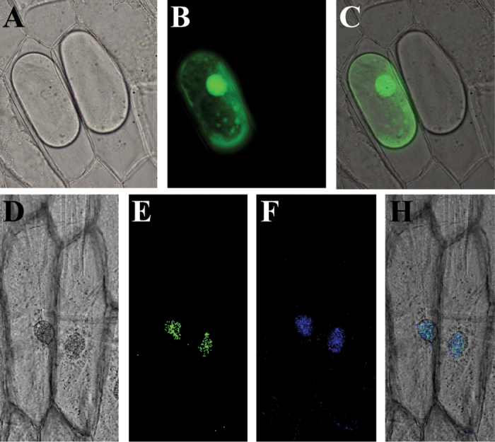
Fig. 2.
Subcellular localization of SlDREB in onion epidermal cells. GFP and SlDREB–GFP fused constructs were expressed transiently in onion epidermal cells. Bright-field images (A, D), GFP fluorescent images (B, E), DAPI image (F), and merged images (C, H) of representative cells transformed with GFP (A–C) or the SlDREB–GFP fusion protein (D–G) are shown. (This figure is available in colour at JXB online.)

