Figure 1.
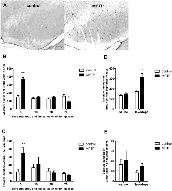
Histological analysis and quantification of the absolute numbers of BrdU+cells in the substantia nigra pars reticulata (SNpr) and compacta (SNpc) at different time points following BrdU and first saline or MPTP injection. Data are expressed as mean +/− S.E.M. A: BrdU-positive cells in the SN of healthy control animals (control) versus MPTP-treated animals (MPTP). Representative coronal 40μm sections at day 3 after BrdU administration and first saline (control) or MPTP injection are given. Scale bar 100μm. B: In short term groups (3 days) MPTP induced a significant increase in the number of BrdU+ cells in the SNpr compared to saline-treated controls. In the 70 days-group MPTP caused a significant decrease in the numbers of BrdU+ cells compared to saline-treated controls. *p < 0.05, **p < 0.001 versus corresponding control (n= 5; LSD post hoc test). C: Only in short term groups (3 days) MPTP induced a significant increase in the number of BrdU+ cells compared to saline-treated controls in the SNpc. **p < 0.001 versus corresponding control (n= 5). D: In the SNpr there was a significant interaction effect between MPTP-treatment (MPTP versus control) and levodopa-treatment (L-Dopa versus saline), as levodopa treatment in addition to MPTP-treatment (MPTP + L-Dopa) increased the number of BrdU+ cells compared to saline-treated controls (control, saline), MPTP and saline treated mice (MPTP, saline) and levodopa-treated mice. *p < 0.05 versus other groups (n ≥ 5; LSD post hoc test). E: There were no effects of levodopa-treatment or MPTP on the number of BrdU+ cells in the SNpc.

