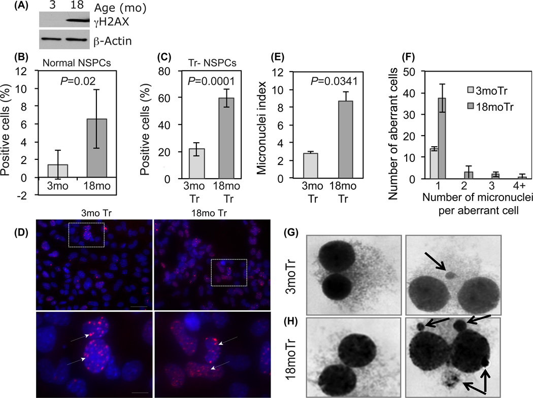
Figure 3. Age dependent activation of γH2AX expression and increased micronuclei formation.
(A) Western blot showing differential γH2AX protein content in normal 3 and 18mo NSPCs. (B) Quantification of γH2AX immunopositivity in cultured normal 3mo and 18mo NSPCs. (C) Quantification of γH2AX foci in 3mo and 18mo Tr-NSPCs. (D) Representative photomicrographs demonstrating detection of γH2AX foci in 3mo and 18mo Tr-NSPCs. Bottom row- higher magnification of areas designated above (arrows indicate foci). (E) Percentage of binucleate cells containing micronuclei (“Micronuclei index”) was significantly greater in 18mo compared to 3mo Tr-NSPCs (8.7% vs 2.8%, p=0.035) [pooled from a 1000 cells counted for each age in two separate experiments]. (F) Frequency of single or multiple micronuclei in aberrant binucleate cells by age. (G, H) Appearance of binucleate cells without micronuclei (left panel) or with micronuclei (right panel, arrows) for 3mo and 18mo Tr-NSPCs, respectively. Multiple micronuclei (right sided panel of 3H) were only detected in 18mo Tr-NSPCs.

