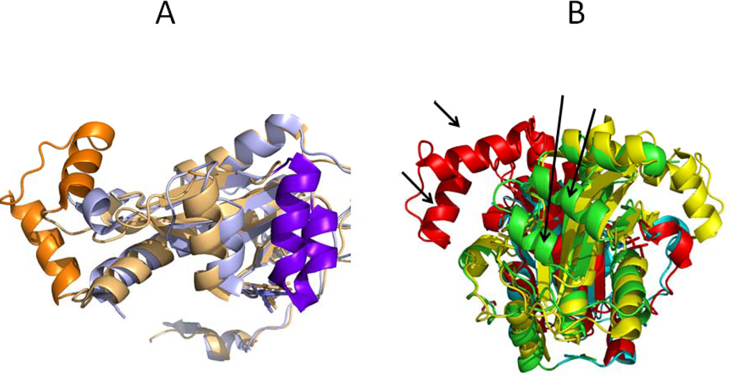
FIGURE 5. Structural comparison of the RdxA and FrxA structures.
A. The RdxA monomer is in blue and the FrxA monomer in gold. The α-helices which are only visible in one of the structures are highlighted in darker colours. B. The two chains o the RdxA dimer are depicted in cyan and green. The two subunits of the biologically relevant FrxA dimer are in red and yellow. The arrows point to the two helices in RdxA not seen in FrxA and to the two helices in FxrA not seen in RdxA

