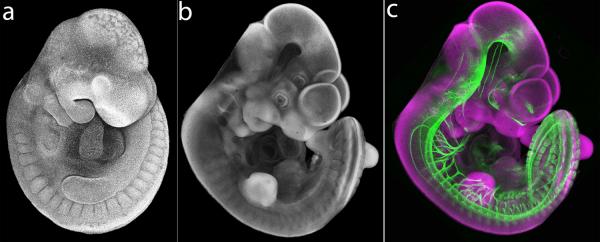
Figure 5.
Comparison of uncleared vs. cleared specimens. (a) Lateral view of uncleared DAPI-stained E10.5 mouse embryo. Tissues in physiological buffer are opaque and appear solid. (b) Lateral view of BABB-cleared DAPI-stained E10.75 mouse embryo. Exterior morphology of cleared specimen is evident, but embryo appears transparent and topological features from the far side of the embryo such as eye and forebrain vesicle are visible. (c) Lateral view of BABB-cleared DAPI-stained E10.75 mouse embryo immunostained for NCAM1. Nuclear DAPI signal is rendered in magenta for optimal contrast with green neuronal antibody staining.

