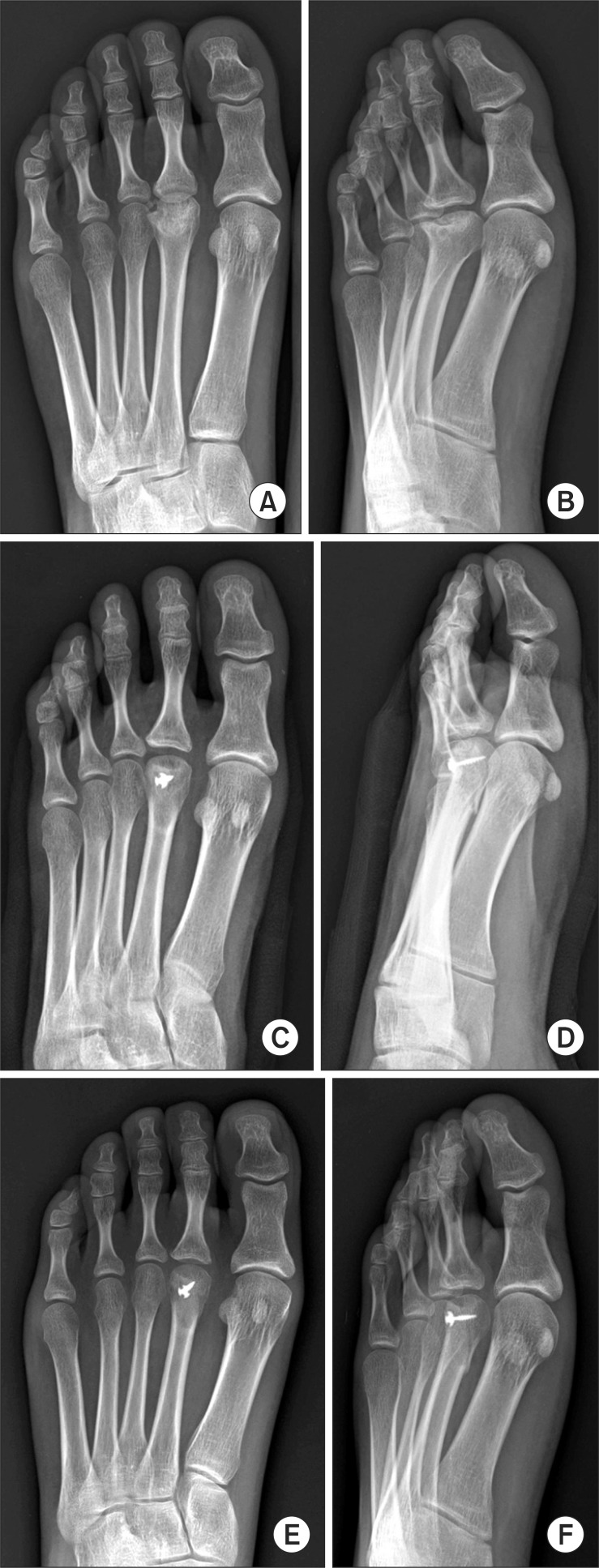
Fig. 2.
A 22-year-old woman with Freiberg's disease (case 16) that was classified as Smillie stage II. (A, B) Preoperative radiography shows that central portion begins to sink into the head, altering the contour of the articular surface. (C, D) Through dorsal closing wedge osteotomy with a screw fixation, the metatarsal was shortened and the plantar part of the metatarsal head was rotated. (E, F) Four weeks later after operation, the bridging trabeculae across the osteotomy site emerged which is considered as radiographic union.

