Figure 4.
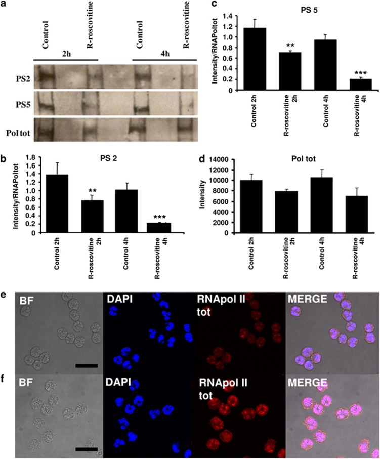
R-roscovitine inhibits RNApol II phosphorylation on serine 2 and serine 5. (a) Western blotting for RNApol II total (Pol tot), phosphorylated on serine 2 (PS2) and phosphorylated on serine 5 (PS5) after 2 and 4 h in control or R-roscovitine (20 μM) treated neutrophils. Lysates were run on 4% acrylamide gels with blots shown representative of three experiments. Densitometry (n=3, mean±S.E.M.) for serine 2 (b) and serine 5 (c), normalised to total RNApol II levels (d). Statistical significance compared to relevant time-point control where P<0.01 is shown as ** and P<0.001 *** by ANOVA with post hoc multivariate analysis by Student's Newman–Keuls (with a 95% confidence interval). Confocal microscopy of RNApol II (total) in neutrophils either unstimulated (e) or stimulated with LPS (100 ng/ml) (f) by indirect immunofluorescence. Panels show transmitted light/bright field (BF), DAPI nuclear staining (blue), RNApol II (total) staining (red), final panel shows merged image (MERGE) demonstrating nuclear colocalisation (pink). Size bar represents 10 μm scale. Leica SP confocal microscope × 630 oil immersion. Representative images are shown from n=3 experiments

