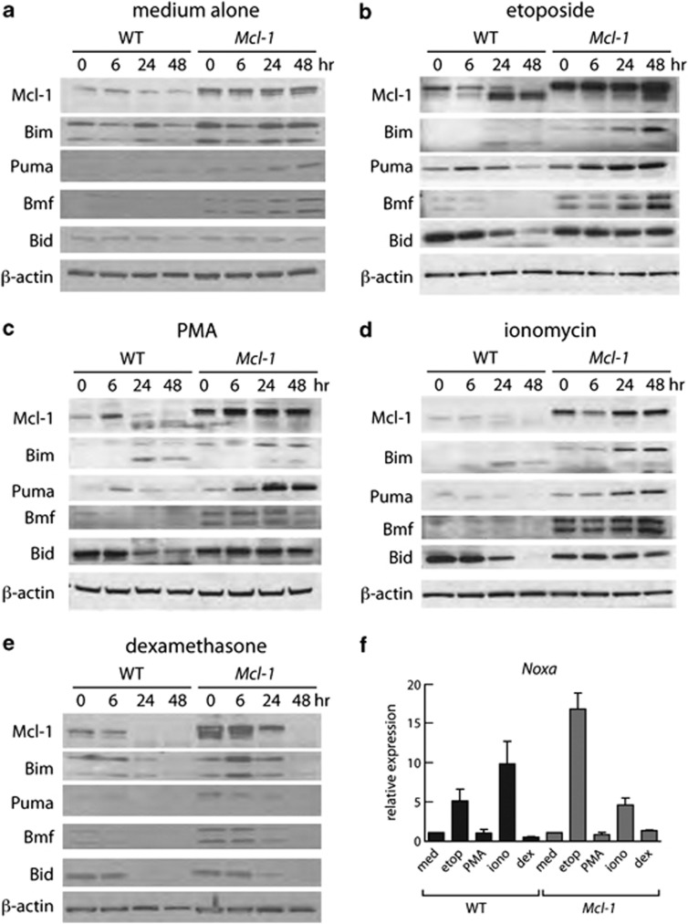
Figure 2.
Mcl-1 levels correlate with thymocyte viability. (a– e) Western blot analysis (20 μg protein) of lysates of thymocytes from WT or Mcl-1tg mice cultured in vitro for indicated times with (a) medium alone, (b) 1 μg/ml etoposide, (c) 10 ng/ml PMA, (d) 1 μg/ml ionomycin or (e) 1 μM dexamethasone. Data are representative of three independent experiments each performed with thymocytes from one mouse of each genotype. Supplementary Table S1 shows quantification of protein levels. (f) Q-PCR analysis of Noxa mRNA expression after culture for 6 h under the conditions described in (a– e). The data are expressed relative to β-actin and the untreated sample for the same genotype. Bim was the only other gene tested that showed significant transcriptional activation at 6 h, and only in Mcl-1tg thymocytes in response to dexamethasone (∼4-fold) (data not shown)

