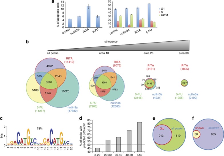
Figure 1.
Characterization of the global chromatin occupancy by p53 upon its activation by nutlin3a, RITA and 5-FU. (a) Induction of apoptosis upon nutlin3a, RITA and 5-FU treatment of MCF7 cells was detected by fluorescence-activated cell sorting (FACS) of annexin V-stained cells (left) and cell-cycle profiles were assessed by FACS of PI-stained cells (right). (b) Venn diagrams were obtained by intersection of all p53-bound DNA fragments (ChIP-Seq peaks, P<0.05) obtained from MCF7 cells treated with nutlin3a-, 5-FU- and RITA and filtering according to the area. (c) The p53 consensus binding motif was identified de novo by analyzing the sequences of 500 randomly selected peaks common for all the three treatments using the program MEME (d). The fraction of peaks containing the p53 consensus site increased along with increased stringency of peak selection. (e) A Venn diagram demonstrates the proportion of p53-bound fragments located within ±10 kb of the TSS (913 out of 2432). (f) A Venn diagram shows the proportion of peaks located in the vicinity of known and unknown p53-target genes bound within ±10 kb of TSS upon all three treatments. See also Supplementary Figure S1, Supplementary Figure S2 and Supplementary Tables S1–S4

