Figure 5.
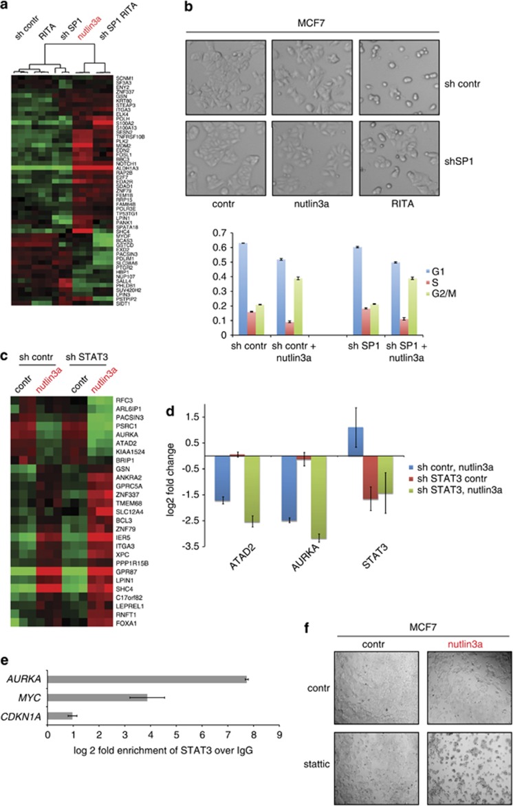
Identification of Sp1 and STAT3 as important modulators of p53 transcriptional activity. (a) The heatmap shows the results of gene expression analysis of RITA-treated MCF7 cells in which Sp1 was stably depleted by short hairpin RNA (shRNA). (b) Sp1 depletion attenuated RITA-induced apoptosis, but did not affect nutlin3a-induced growth arrest, as assessed by microscopy analysis (upper panel) and FACS of PI-stained cells (lower panel). (c) The heatmap shows the results of gene expression analysis of nutlin3a-treated MCF7 cells in which STAT3 was stably depleted by means of shRNA. (d) Depletion of STAT3 by shRNA enhances p53-mediated repression of AURKA and ATAD2 as assessed by qPCR in MCF7 cells treated with nutlin3a. (e) STAT3 ChIP in MCF7 cells demonstrated the binding of STAT3 to the promoter of AURKA. CDKN1A served as a negative and MYC as a positive control for STAT3 chromatin binding. (f) Inhibition of STAT3 by stattic facilitated the growth-suppression effect of nutlin3a as assessed by light microscopy analysis of the MCF7 after 48 h of treatment. See also Supplementary Figure S5

