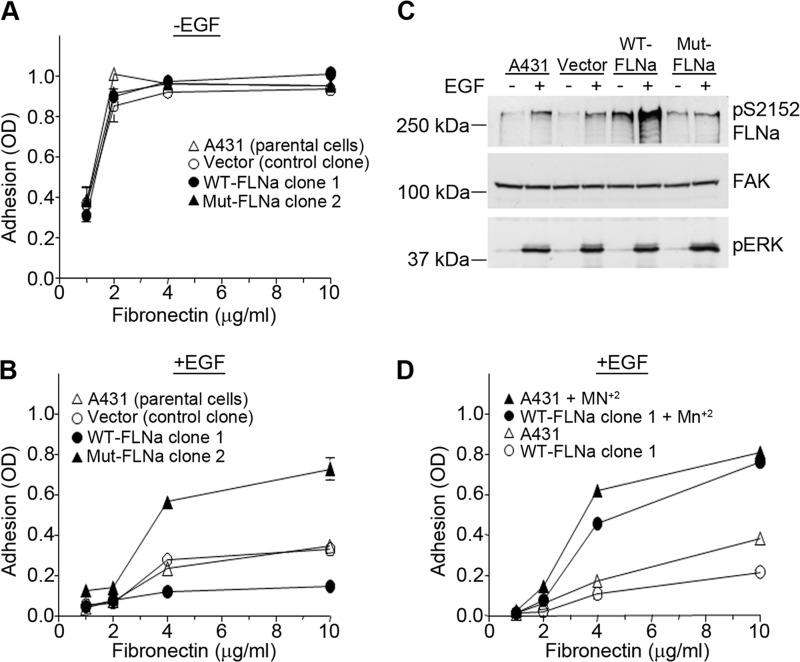
FIGURE 8.
EGF modulates a5β1 integrin affinity through phosphorylation of FLNa. A and B, A431 parental cells, a vector control clone, WT-FLNa clone 1, and Mut-FLNa clone 2 were allowed to adhere to wells coated with increasing concentrations of fibronectin in the absence (A) or presence (B) of 3 nm EGF. Cell adhesion assays were performed as described in the legend to Fig. 1. Error bars indicate S.D. of triplicate samples for one of three representative experiments. C, A431 parental cells, a clone containing only the pcDNA3 vector, WT-FLNa clone 1, and Mut-FLNa clone 2 were treated with EGF (3 nm) for 20 min. Cell lysates were prepared, and phosphorylated FLNa was visualized by Western blotting using an antibody to phosphorylated FLNa. FAK was used as a loading control, and phospho-ERK (pERK) served as a positive control for EGF treatment. Western blots are representative of three separate experiments. pS2152 FLNa, phospho-Ser-2152 FLNa. D, A431 parental cells and WT-FLNa clone 1 were pretreated for 1 h at room temperature with or without 0.5 mm MnCl2. Cells were then seeded onto wells coated with increasing concentrations of fibronectin in the presence of 3 nm EGF. Cell adhesion was assessed as described in the legend to Fig. 1. Error bars indicate S.D. of triplicate samples for one of three representative experiments.

