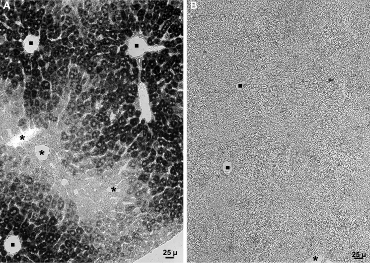
FIGURE 3.
In situ hybridization showing Sepp1 mRNA (dark stain) in mouse liver. Portal veins are indicated by filled squares and central veins by asterisks. Mice had been fed a diet supplemented with 0.25 mg of selenium/kg. A depicts liver from a C57BL/6 mouse and B depicts liver from a congenic Sepp1−/− mouse.

