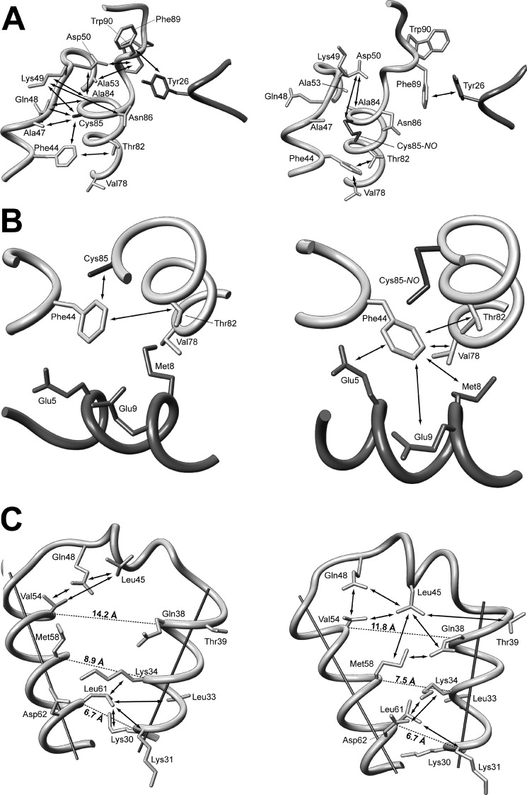
FIGURE 4.
Fragments of high resolution three-dimensional structures of reduced (left) and S-nitrosylated (right) variants of human S100A1 protein. A, the part of the structure showing the C terminus, hinge region, and N-terminal Ca2+-binding loop. B, differences in the contacts of Phe44 aromatic side chain with the C-terminal part of helix IV and central part of helix I′. C, maps of contacts observed between residues from helix II, helix III, and the hinge region.

