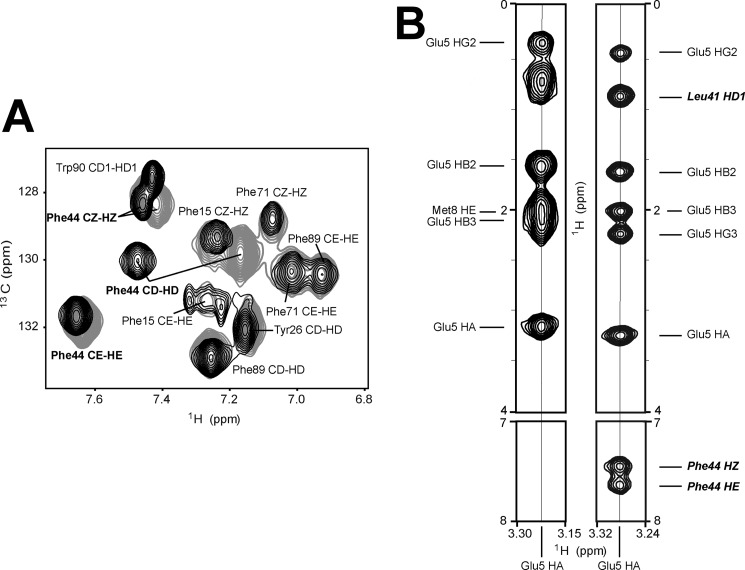
FIGURE 5.
NMR data on aromatic side chains in reduced and S-nitrosylated variants of apo-S100A1 protein. A, overlay of two-dimensional aromatic 1H-13C HSQC spectra acquired for apo-S100A1 protein in reduced (black) and S-nitrosylated (gray) form. B, two-dimensional 1H-1H planes of three-dimensional 13C-edited NOESY-HSQC spectra taken at the frequency corresponding to 1Hα Glu5. 1Hα Glu5-1Hϵ Phe44 and 1Hα Glu5-1Hz Phe44 are clearly observed in the case of apo-S100A1-NO.

