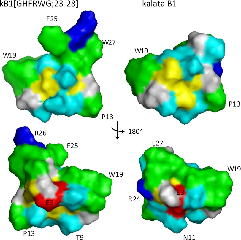
FIGURE 6.
Surface representation of kB1(GHFRWG;23–28) (left) and kalata B1 (right). The only major differences are observed in the grafted loop 6, i.e. residues 23–28. Hydrophobic residues are highlighted in green, positively charged in blue, negatively charged in red, cysteines in yellow, and polar in cyan. The top views have similar orientation as the ribbon models in Fig. 5. The residues F25, R26, and W27 in kB1(GHFRWG;23–28) are part of the melanocortin pharmacophore grafted into loop 6 of kalata B1.

