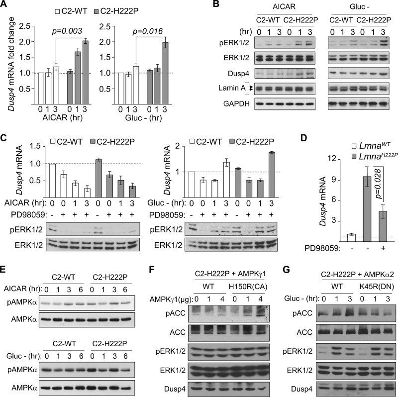
FIGURE 2.
Dusp4 expression is enhanced in response to energy deficit in C2C12 stably expressing H222P-lamin A. A, qPCR analysis of Dusp4 mRNA 0, 1, and 3 h after AICAR treatment (left panel) or glucose deprivation (Gluc −, right panel) in C2C12 cells stably expressing FLAG-tagged WT lamin A (C2-WT) or H222P-lamin A (C2-H222P) (n = 3 experiments). B, Western blot of protein extracts 0, 1, and 3 h after AICAR treatment (left panel) or glucose deprivation (Gluc −; right panel) in C2C12 stably expressing C2-WT or C2-H222P. The blots were probed with antibodies against phospho-ERK1/2 (pERK1/2), total ERK1/2 (ERK1/2), DUSP4, and lamin A. Two bands in the lamin A blot represent the FLAG-tagged and endogenous proteins. A representative blot is shown from five experiments. C, qPCR analysis of Dusp4 mRNA and representative Western blot of pERK1/2 and ERK1/2 in C2-WT and C2-H222P subjected to 30 min of pretreatment with PD98059 followed by PD98059 and AICAR co-treatment (left panel) or glucose deprivation (right panel) for 0, 1, and 3 h. Dusp4 mRNA values are presented as fold change over untreated C2-WT. Untreated and treated with PD98059 are denoted by − and +, respectively (n = 3 experiments). D, qPCR analysis of Dusp4 mRNA in hearts of LmnaH222P/H222P (LmnaH222P) and WT (LmnaWT) mice subjected to 4 weeks of treatment with Me2SO or PD98059. Dusp4 mRNA values are presented as fold change over WT. Treatment with or without PD98059 (Me2SO given in place of PD98059) are denoted by − and +, respectively (n = 4). E, Western blot analysis of phospho-AMPK and AMPK in C2-WT and C2-H222P treated with either AICAR (top panels) or subjected to glucose deprivation (Gluc −; bottom panels) for 0, 1, 3, and 6 h. A representative blot from three experiments is shown. F, Western blot analysis of phospho-ACC, ACC, phospho-ERK1/2, ERK1/2, and DUSP4 in C2-H222P transiently transfected with increasing concentration of WT AMPKγ1 or H150R constitutively active (CA) AMPKγ1 point mutant expression vector. A representative blot from two experiments is shown. G, Western blot analysis of phospho-ACC, ACC, phospho-ERK1/2, ERK1/2, and DUSP4 in C2-H222P transiently transfected with WT AMPKα2 or K45R dominant negative (DN) AMPKα2 point mutant expression vector subjected to 0, 1, and 3 h of glucose deprivation. A representative blot from two experiments is shown.

