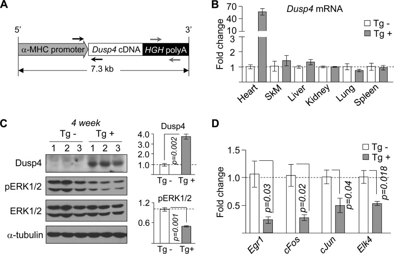
FIGURE 3.
Cardiac-selective overexpression of Dusp4 reduces ERK1/2 signaling. A, schematic diagram of targeting vector used to generate Dusp4 transgenic mice. α-MHC denotes α-myosin heavy chain, and HGH poly(A) denotes human growth hormone polyadenylation signal. The black and gray arrows indicate annealing positions of genotyping primers. B, qPCR analysis of Dusp4 mRNA in various tissues (SkM denotes skeletal muscle) from 4-week-old Dusp4 Tg+ and Tg− littermates (n = 3 samples). C, Western blot of phosphorylated ERK1/2 (pERK1/2), total ERK1/2 (ERK1/2), DUSP4, and α-tubulin in hearts of 4-week-old Dusp4 Tg+ and Tg− littermates (left panel) (n = 3 samples). The numbers above the blots denote individual samples. Quantification of DUSP4 and pERK1/2 normalized to α-tubulin and ERK1/2, respectively, is presented as fold change over WT (right panel). D, qPCR analysis of expression of genes downstream of ERK1/2 in 4-week-old Dusp4 Tg+ and Tg− littermates (n = 3 samples).

