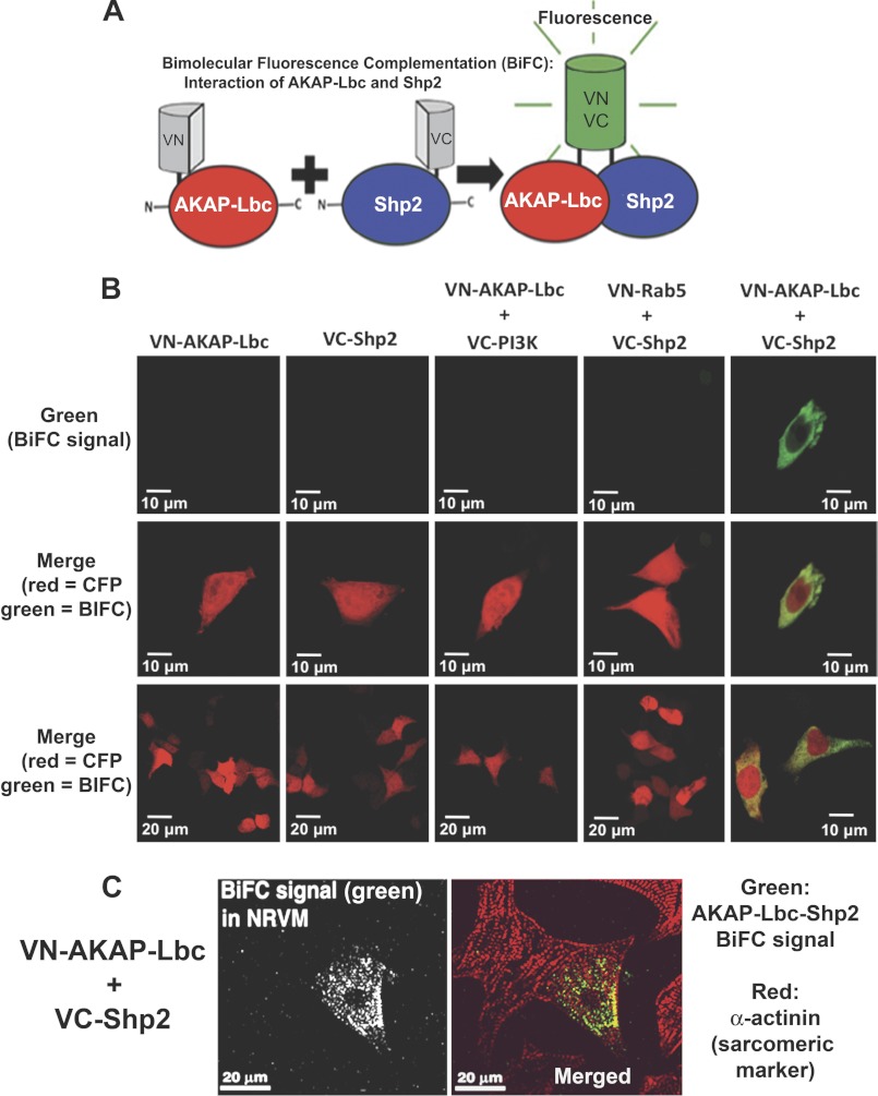
FIGURE 2.
Visualization of AKAP-Lbc-Shp2 interaction inside cells by BiFC. A, schematic diagram illustrates the BiFC assay. The Venus (green) fluorescent protein is split into two nonfluorescent halves which are fused to AKAP-Lbc and Shp2. Specific protein-protein interaction results in a functional Venus (green) fluorescent protein. B, HEK293 cells were co-transfected with VN-AKAP-Lbc and VC-Shp2 and CFP (pseudo-colored red), as a marker for transfected cells. Cells were fixed 20 h after transfection and imaged. No BiFC (green) fluorescence is observed in cells expressing either VN-AKAP-Lbc or VC-Shp2 alone or when VN-AKAP-Lbc is co-expressed with a noninteracting control VC-protein (VC-PI3K). Similarly, no BiFC (green) fluorescence is observed when VC-Shp2 is co-expressed with a control VN-protein (VN-Rab5), whereas specific interaction of VC-Shp2 with VN-AKAP-Lbc results in fluorescence. Equal protein expression was determined by Western blotting (data not shown). C, neonatal rat ventricular cardiac myocytes were electroporated for expression of VN-AKAP-Lbc and VC-Shp2. After 48-h treatment with phenylephrine (10 μm), cells were fixed, permeabilized, and immunostained for the sarcomeric marker protein α-actinin (red in merged image). BiFC signal is shown in green in the merged image.

